Toggle filters
    Photos
    Expand filters
    • Search by Image
    Reset color
    Toggle filters
    Photos
    Expand filters
    • Search by Image
    Reset color

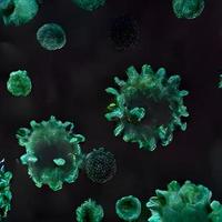
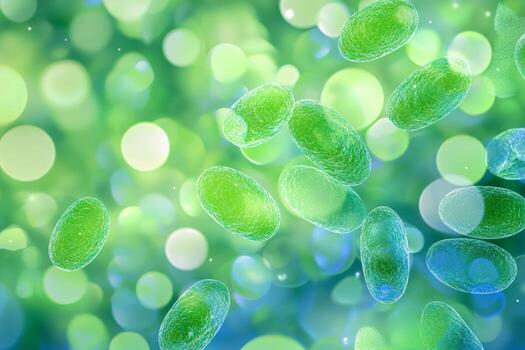
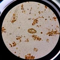
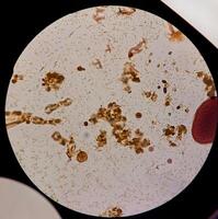
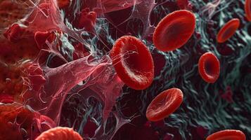
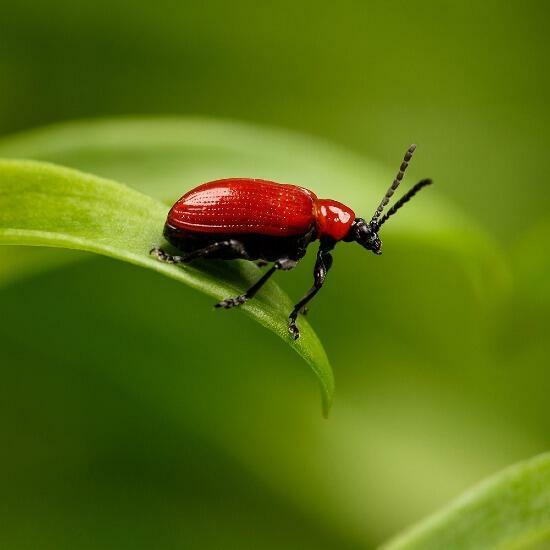
    Cellular Microorganism Photos & Images (Page 4)

     - 575 high resolution, royalty free stock photos and pictures matching Cellular Microorganism

    • Close up of a scientific 3D illustration showing virus particles in detail with a bright and colorful microscopic environment photo
    • Biological cells under a microscope. The microorganisms microbes, bacteria, protein. photo
    • Biological cells under a microscope. The microorganisms microbes, bacteria, protein. photo
    • Virus particles floating in a dark space with a blurred background showcasing structure and detail photo
    • Virus structure with spikes shown in detail against a blurred background highlighting the importance of microscopic analysis photo
    • Close-Up of Bacteria Cell Structure with Detailed Microbial Features photo
    • Colorful virus particles are shown interacting with cells in a vibrant biological setting at a microscopic level photo
    • Bacteria exhibit vibrant blue colors while moving through a dark microscopic environment, highlighting their structure photo
    • A virus under a microscope showcases intricate details in a vibrant blue background, illustrating biological complexity photo
    • A vibrant bacterium stands out among similar microbes, showcasing its unique shape and intricate details photo
    • Microscopic view of bacteria drifting in water, showcasing their unique shapes and structures in a dark environment photo
    • covid-19, coronavirus outbreak, virus floating in a cellular environment , coronaviruses influenza background, viral disease epidemic, 3D rendering of virus, organism illustration, virus seen micro photo
    • covid-19, coronavirus outbreak, virus floating in a cellular environment , coronaviruses influenza background, viral disease epidemic, 3D rendering of virus, organism illustration, virus seen micro photo
    • covid-19, coronavirus outbreak, virus floating in a cellular environment , coronaviruses influenza background, viral disease epidemic, 3D rendering of virus, organism illustration, virus seen micro photo
    • covid-19, coronavirus outbreak, virus floating in a cellular environment , coronaviruses influenza background, viral disease epidemic, 3D rendering of virus, organism illustration, virus seen micro photo
    • covid-19, coronavirus outbreak, virus floating in a cellular environment , coronaviruses influenza background, viral disease epidemic, 3D rendering of virus, organism illustration, virus seen micro photo
    • covid-19, coronavirus outbreak, virus floating in a cellular environment , coronaviruses influenza background, viral disease epidemic, 3D rendering of virus, organism illustration, virus seen micro photo
    • covid-19, coronavirus outbreak, virus floating in a cellular environment , coronaviruses influenza background, viral disease epidemic, 3D rendering of virus, organism illustration, virus seen micro photo
    • Blood sample for Meningitis viral panel test with laboratory background. photo
    • Colorful Microscopic View of Bacteria Under Magnification for Scientific Use photo
    • Microscopic view of bacteria cells floating in a body photo
    • Close-up view of bacteria under a microscope with soft lighting showcasing detailed cellular structures photo
    • Microscopic view of multiplying bacteria and viruses with neon green and blue lighting in a minimalist setting photo
    • Microscopic depiction of bacteria and viruses multiplying in vibrant neon green and blue lighting photo
    • Close-up view of various microorganisms under a microscope, showcasing bacteria and other tiny particles. photo
    • Close-up view of various microorganisms under a microscope, showcasing bacteria and other tiny particles. photo
    • Close-up view of various microorganisms under a microscope, showcasing bacteria and other tiny particles. photo
    • This visual showcases bacteria displaying vibrant colors floating in a dynamic environment, illuminated by various shades of purple and blue, highlighting intricate details of their structure. photo
    • A cluster of vibrant bacteria is seen actively reproducing in a deep aquatic setting, illuminated by soft violet and blue light, creating a striking contrast against the dark backdrop. photo
    • Bacteria and microorganisms of various shapes and colors populate a vibrant human gut, showcasing the intricate and dynamic ecosystem. This microscopic view highlights the diversity of life inside. photo
    • Surreal microscopic view of bloodstream with cells and pathogens photo
    • Colorful microscopic world of bacteria photo
    • Microscopic exploration of cell structure photo
    • Organic microbial tentacle background, 3d rendering. photo
    • Detailed 3d illustration of bacteria in microscopic view photo
    • Close-Up of Microscope With Red Light. AI-generated photo
    • A computergenerated image depicts a virus floating in water photo
    • An illustration showing cells suspended in liquid photo
    • A computer generated image shows a virus moving through a cell photo
    • An isolated beta cell in the pancreas showing its intricate cellular structure and organelles photo
    • An allergic reaction captured at the cellular level with mast cells releasing histamine and causing nearby vessels to dilate photo
    • The folding of proteins like a puzzle with each piece fitting perfectly into its designated spot photo
    • An overview of collagen synthesis from transcription of the gene to the final formation of mature collagen fibers photo
    • A confocal microscopy image highlighting the role of intermediate filaments in supporting and stabilizing the cells cytoskeleton during movement photo
    • A phasecontrast microscopy image capturing the flow of cytoplasm and the shifting of cellular components as the cytoskeleton reorganizes during cell movement photo
    • The intricate network of neuronal connections displayed through tered synaptic vesicles at a synapse photo
    • A single Tcell engulfing a bacterium through phagocytosis photo
    • An enlarged view of a cell in metaphase with the chromosomes aligning at the equator of the cell before being pulled apart photo
    • A fluorescence microscopy image showing the release of CO2 as a byproduct of the citric acid cycle in cellular respiration photo
    • Crosssection view of the mitochondria showcasing the arrangement of proton pumps in the membrane photo
    • An artistic rendering of the complex network of molecular chaperones within a cell working together to prevent protein misfolding and maintain cellular function photo
    • A crosssection of a cell membrane showing specific receptors interacting with incoming molecules for endocytosis photo
    • An artistic rendition of clathrin proteins depicting their dynamic and versatile role in cellular processes such as endocytosis photo
    • A colorful fluorescence image of peroxisomes highlighting their crucial role in maintaining cellular homeostasis through fatty acid breakdown photo
    • Cross section of a cell displaying glycogen granules layered between other organelles highlighting their essential role in cellular metabolism photo
    • A closeup of a lysosome filled with various cellular debris and waste products photo
    • Multicolored biochemical pathways illustrating the complexity of cellular metabolism photo
    • High magnification image of cytoplasmic streaming in a protist revealing the intricate network of cytoplasmic threads photo
    • An immunofluorescence image of a cell undergoing cellular respiration with bright spots indicating the active production of energy within mitochondria photo
    • Changes in chloride channel activity have been linked to disorders such as hypertension and heart disease highlighting their importance beyond cellular function photo
    • A fluorescentlabeled image of newly synthesized collagen molecules being transported along microtubules towards the extracellular matrix photo
    • The importance of actin remodeling in immune synapse formation with various microtubule structures visible near the membrane photo
    • A protein structure with a large section missing showing where a section of the protein was cleaved off in posttranslational modification photo
    • An upclose image of a single Golgi apparatus stack showing different regions responsible for different stages of protein processing photo
    • An intricate network of vesicles and proteins involved in exocytosi photo
    • A series of timelapse images showing the dynamic process of differentiation in embryonic stem cells as they gradually acquire specialized features and functions photo
    • A network of nuclear pores in a cell nucleus crucial for maintaining proper cellular function and communication photo
    • A combination of brightfield and fluorescence microscopy highlighting clathrincoated pits as dark spots in a sea of fluorescentlylabeled cellular components photo
    • The impressive ability of the endoplasmic to correctly fold and process a vast amount of proteins for cellular use photo
    • An artistic representation of synovial cells with vibrant colors highlighting their vital role in joint lubrication and protection photo
    • Comparative image of normal and mutated proton pumps demonstrating the importance of functional pumps for cellular processes photo
    • A snapshot of the cellular membrane with numerous receptor proteins protruding and ready to receive extracellular signals photo
    • A highmagnification image of the process of oxidative phosphorylation where oxygen is used to generate ATP in the final step of cellular respiration photo
    • A glimpse of exocytosis captured under electron microscop photo
    • A crosssection of a cell showing the dynamic movement and communication between different signaling molecules and organelles photo
    • An illustration showing the complex interplay between signaling pathways and cellular structures involved in cancer cell invasion photo
    • A highpowered image of a DNA damage response protein activated in response to mutations or other forms of damage during the cell cycle photo
    • A of lysosomes surrounding a damaged organelle ready to break it down photo
    • A timelapse image showing the continuous activity of lysosomes constantly degrading and recycling cellular waste photo
    • An upclose view of chondrocytes in the cartilage revealing their ovalshaped nuclei and pale blue cytoplasm photo
    • Microscopic view of accumulated lipofuscin granules within a cellular vacuole a common characteristic of senescent cells photo
    • Enhanced image of telomerase extending the telomere sequence a crucial process in maintaining cellular lifespan photo
    • A microscopic view of the enzyme complexes encoding the citric acid cycle the second step in cellular respiration photo
    • The interior of a mast cell filled with granules containing histamine and other chemical messengers photo
    • A buildup of oxidative stress and damaged DNA within a senescent cell leading to cellular dysfunction photo
    • An electron micrograph revealing the dynamic movement of microfilaments as they aid in cellular locomotion photo
    • The cellular machinery of a contracting cardiac muscle cell with myofibrils and sarcomeres clearly visible photo
    • The engulfment of damaged or dead cells by a macrophage through phagocytosis to clear out cellular debris photo
    • A microscopic view of hematopoietic stem cells floating in a sea of red cells ready to undergo differentiation photo
    • Multiple cells with varying types of CAMs demonstrating their diverse functions in regulating cellular adhesion and signaling photo
    • Visualization of signaling events mediated by CAMs regulating cellular processes such as proliferation differentiation and apoptosis photo
    • Exocytotic fusion of vesicles creating a portal for molecules to exit the cel photo
    • The importance of proper protein folding as misfolded proteins can have detrimental effects on cellular function photo
    • A microscopic view of a cell in the prophase stage with the nuclear membrane disintegrating and the chromosomes condensing photo
    • The endoplasmic as a dynamic environment constantly folding and modifying proteins to meet cellular demands photo
    • A highresolution image of the intricate network of actin filaments involved in the process of phagocytosi photo
    • An electron microscopy image of RNA polymerase binding to a DNA template with the help of transcription factors photo
    • A microscopic image of a developing erythrocyte in the midst of hemoglobin synthesis showcasing the vibrant red color photo
    • At the cellular level protein fibers of the ECM act as scaffolding for cells to attach and organize themselves photo
    Log In to Vecteezy
    or
    Reset Password
    Back to Log In →
    Single Sign-on

    Log in with your team's identity provider:

    ← Back to Log In